

Dental cavities are one of the most common worldwide dental problems, affecting millions yearly. Cavities are caused by decay and bacteria that break down the tooth’s enamel, eventually forming a hole or a cavity, which requires a dental filling to fix. These holes can cause a lot of pain and discomfort and, if left untreated, can lead to more serious dental problems.
Dental cavities are not always visible to the naked eye, so dentists recommend annual X-rays. Dental X-rays provide additional information beyond what is visible during a clinical oral examination. These include proximal dental caries (cavities between teeth) and early carious lesions (tiny cavities) that are too small to be detected otherwise. If they are caught early using dental X-rays, cavities can be reversed, preventing more expensive dental procedures from occurring.
This article will explore what a cavity looks like on an X-ray and how to tell what type of treatment you need.
What do Cavities Usually Look Like on an X-Ray?
Cavities on X-rays appear different than those in the mouth because X-rays are in black and white. There are three things to look out for in the mouth when looking at an X-Ray:
- Cavities: Cavities on X-rays are called radiolucencies. Radiolucencies are dark areas on an X-ray, indicating the presence of hollow areas. Since cavities are just holes within your teeth, these radiolucencies are almost always cavities.
- Healthy Teeth: Healthy tooth structures will appear more radiopaque, or in other words, more dense. This means healthy tooth structure will look brighter areas on an X-ray.
- Fillings/Dental Work: A filling will look even more radiopaque than the surrounding tooth structure and is often extremely white (but it depends on the material used). This is because these materials are specifically designed to look extremely different from your regular tooth. That way, your dentist knows what healthy tooth structure is and what is past dental work.
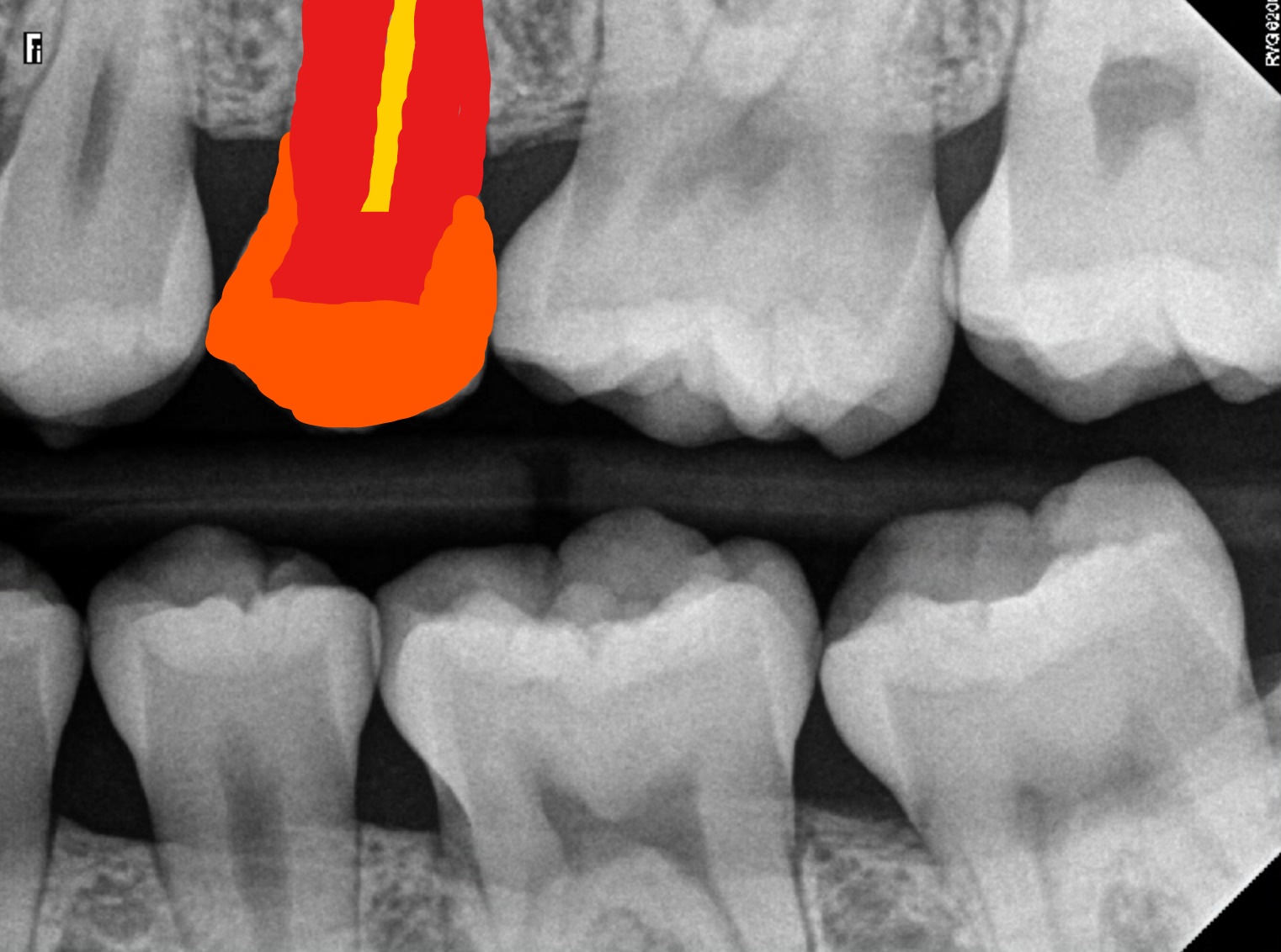
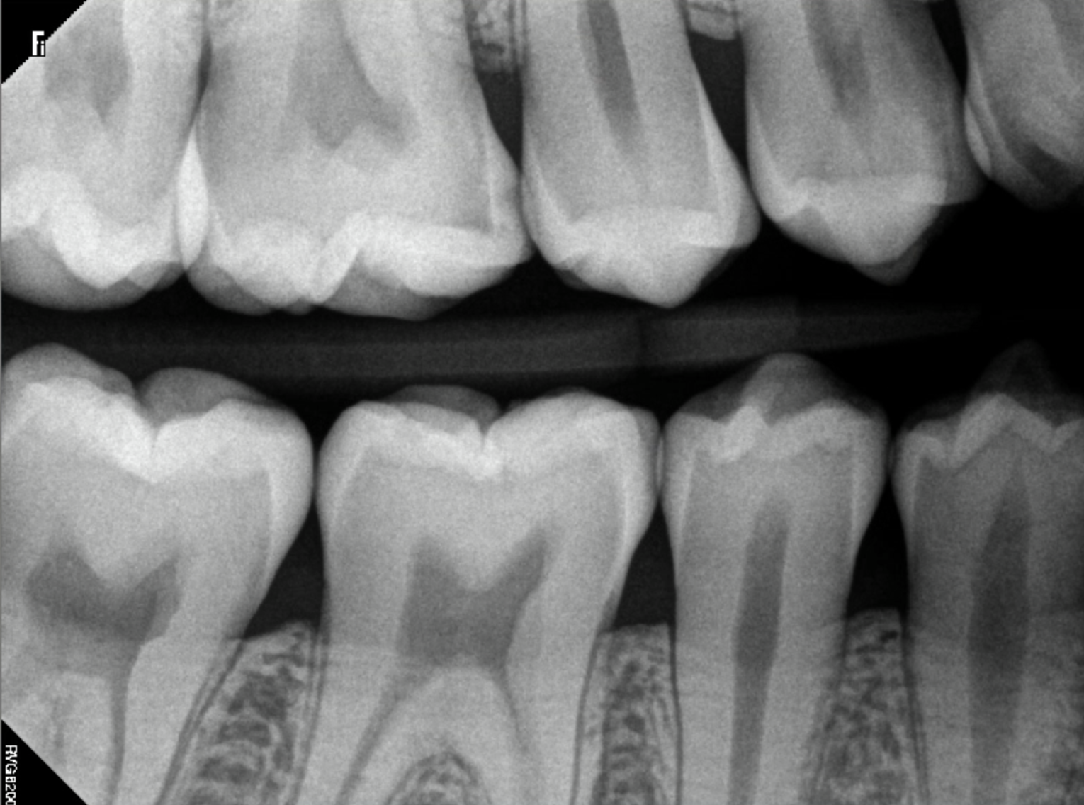
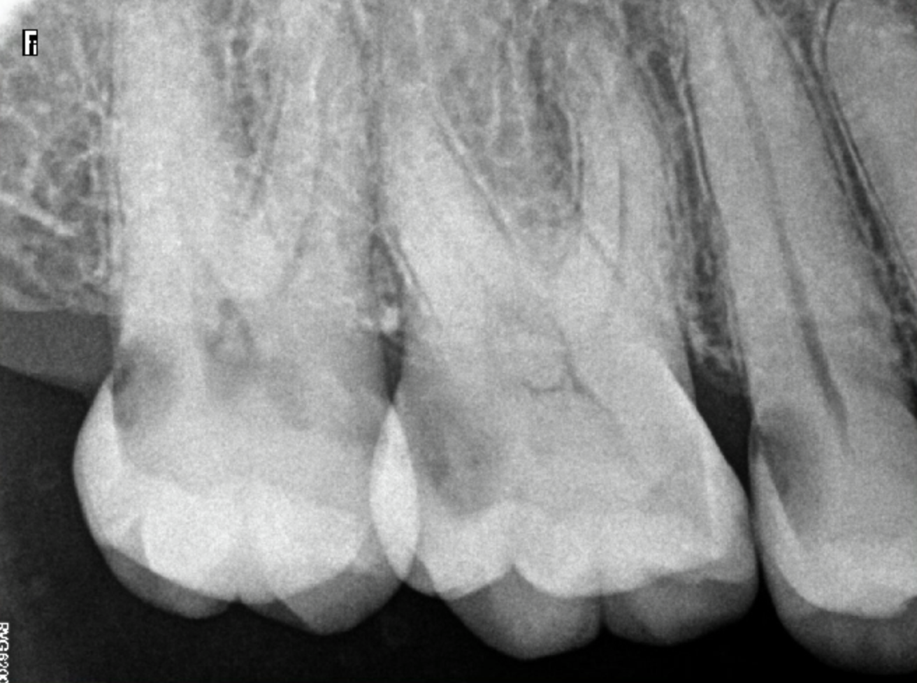
X-Rays of Teeth Without Cavities
Below is a picture of teeth without any cavities for reference. The picture on the left has no fillings whatsoever and is an example of what a fairly healthy tooth will look like on an X-Ray. The outermost layer, the enamel, appears very bright on an X-ray and is the most radiopaque. The middle layer, called dentin, also appears bright but not as much as the enamel. The innermost layer of a tooth, the pulp, is the least dense and appears very dark on an X-Ray. We have colored in this area to make it easier to understand. The pulp is yellow, the dentin is red, and the enamel is orange.
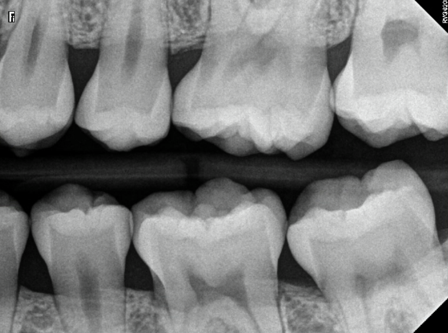
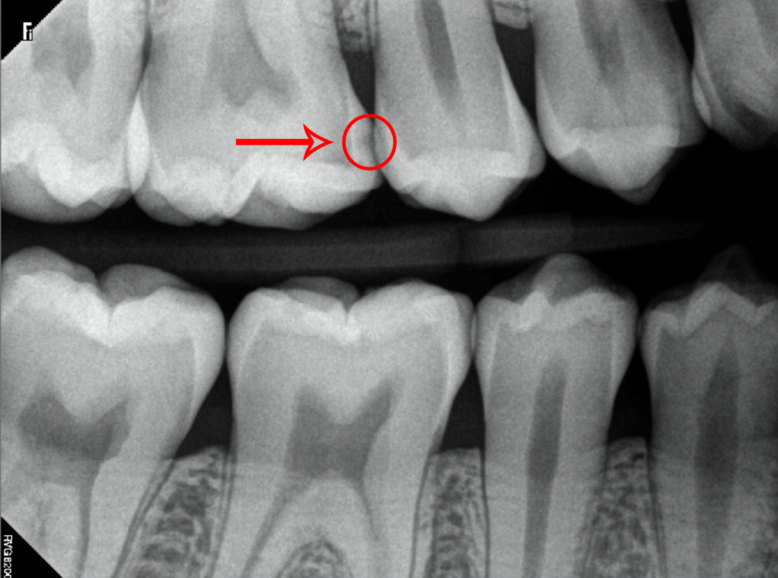
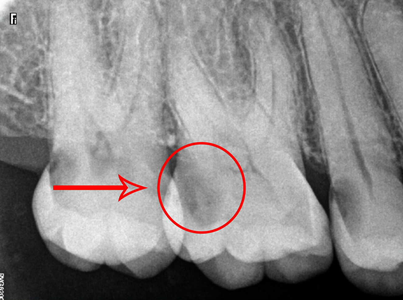
X-Ray of Teeth Under Observation (Mild Cavity)
Teeth under observation are those that have the starting of a cavity forming. However, they have not yet broken past the enamel surface of the teeth and are still in their earliest stages. These cavities can be difficult to spot on an X-Ray, but they usually appear as super tiny and faint radiolucencies. These cavities can be reversed if treated appropriately, and no further damage should occur.
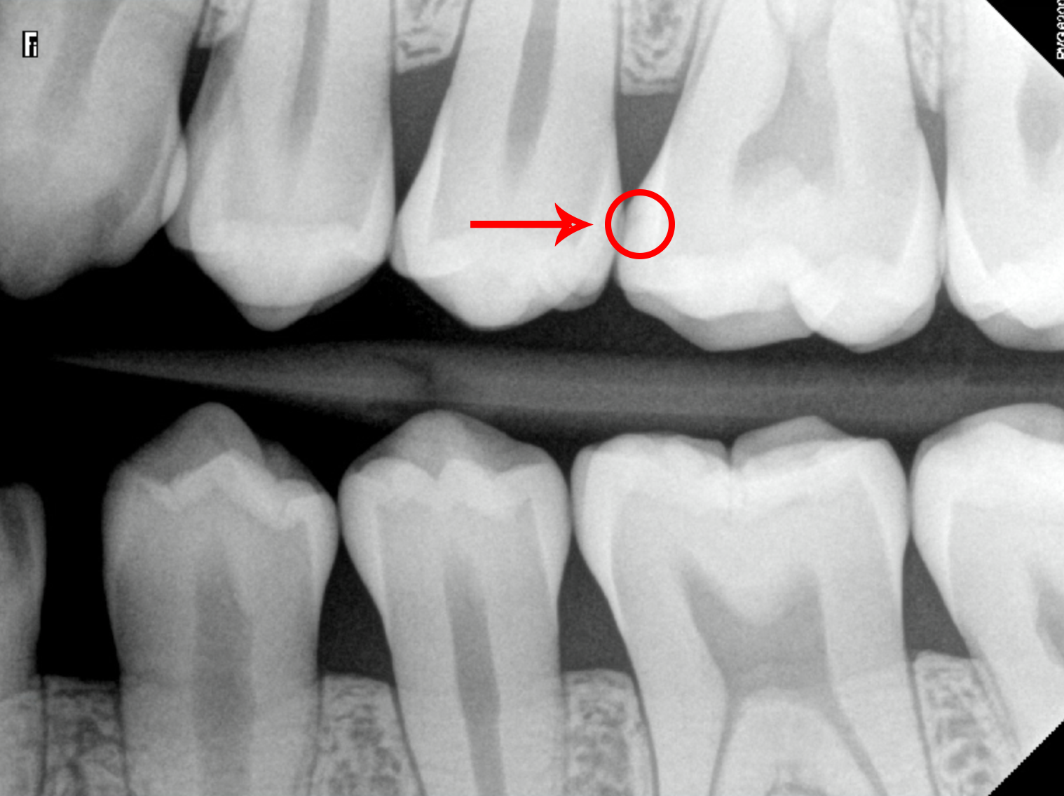
In the case of the image below, the patient’s upper left first molar (tooth number 14 for the American tooth numbering system or 26 for the Canadian tooth numbering system) is under observation and shows a tiny radiolucency that has just begun to form on the mesial surface of the tooth. The mesial surface is the part closest to the center of your mouth and is located between your teeth.
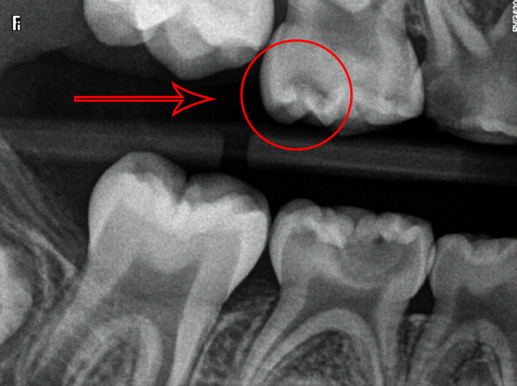
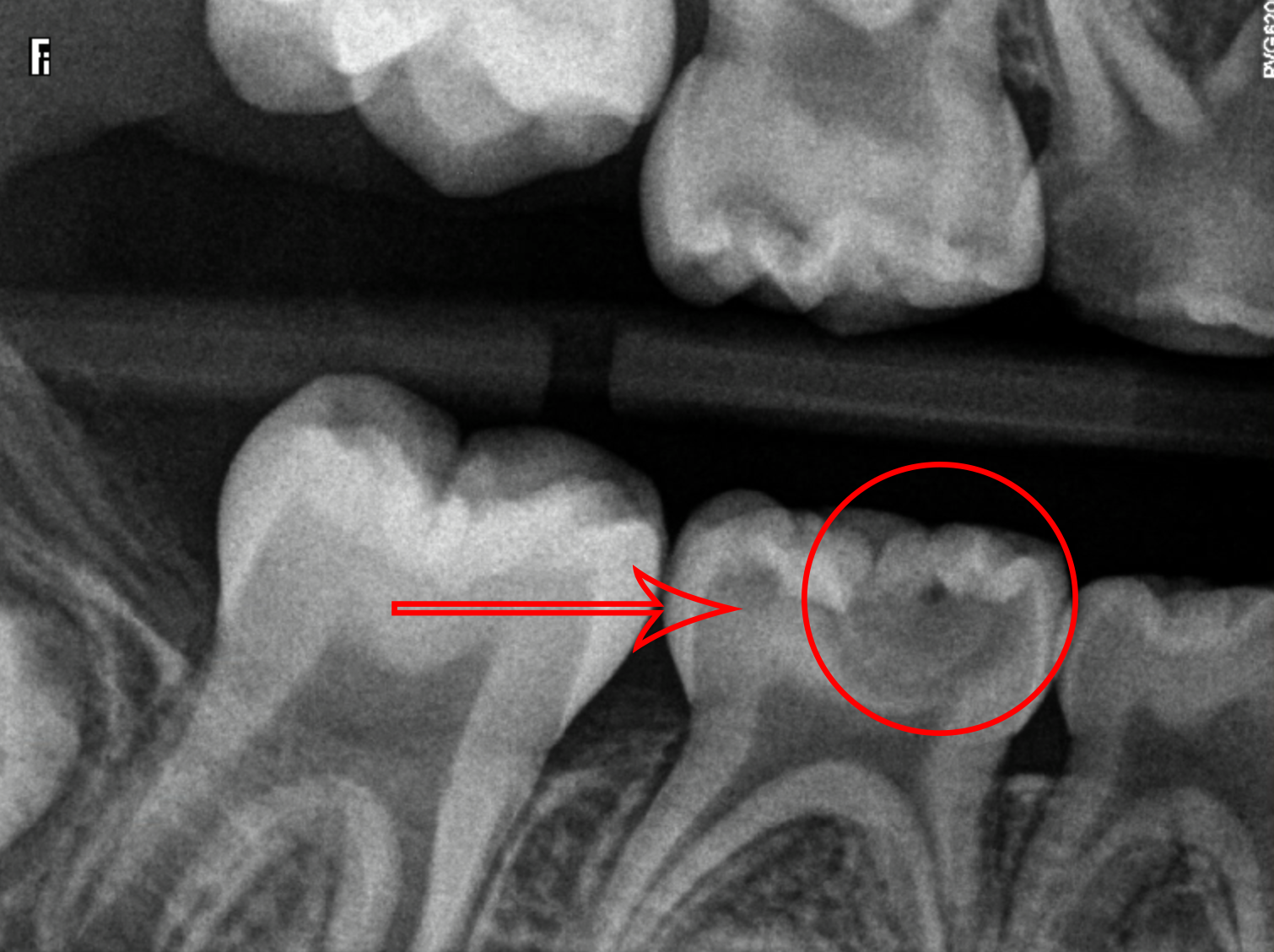
X-Rays of Teeth With Moderate Cavities
When cavities become more severe, they will start to become more visible on X-rays. These cavities will have grown deeper into the tooth, past the enamel and into the dentin, and typically appear as a darker area on an X-Ray. Some cavities may even have “holes” that are more irregular in shape compared to teeth without cavities. These types of cavities will require a dental filling to restore them properly.
In the case of the image below, the patient’s upper right first molar (tooth number 3 for Americans or 16 for Canadians) has a substantially sized cavity visible on the X-ray. This cavity is also on the mesial surface of the tooth and can be more easily seen because it has grown past the enamel layer.
In this image, a child has multiple cavities. We are paying attention to the cavity on their upper right second molar (tooth A for Americans or 55 for Canadians). This cavity can be seen as a darker area than the other teeth and is located on the occlusal surface of the tooth. The occlusal surface is the part of your tooth that you bite down on.
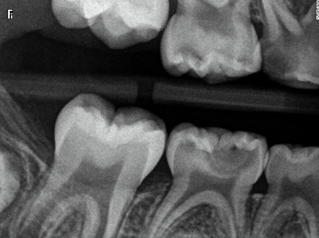
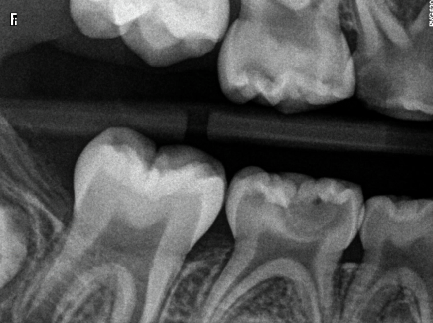
X-Rays of Teeth With Severe Cavities
If cavities are left untreated, they will continue to grow and spread deeper into the tooth. At this point, the cavity may have eaten away the entire tooth structure, past the enamel and dentin and into the pulp, and will appear as a large dark area on an X-Ray. These cavities are extremely painful and will require a root canal or extraction to be treated.
In the case of the image below, the patient has cavities on all their teeth. The cavity we are focusing on is the upper right first molar (tooth number 3 for Americans or 16 for Canadians) which has a severely large cavity. This cavity is on the distal surface of the tooth and can be easily seen because it has eaten away much of the tooth structure.
Looking back at our previous image of the child having multiple cavities, there is also a cavity that has reached the pulp. We are now paying attention to the cavity on their lower right second molar (tooth T for Americans or 85 for Canadians). This occlusal cavity is much more severe and can be seen as a large dark area on the X-Ray where the majority of the tooth structure has been eaten away. For children, a baby root canal or extraction will be recommended.
Conclusion
In conclusion, cavities can range from mild to severe, and the severity of the cavity greatly affects what it looks like on an X-Ray. Cavities that are beginning to form may appear as tiny and faint radiolucencies, while more severe cavities will have eaten away the entire tooth structure and appear much darker on X-Rays. By understanding what cavities look like on X-Rays, dentists can detect and treat them in the early stages and help prevent more severe damage down the line.
Disclaimer
The contents of this website, such as text, graphics, images, and other material are for informational purposes only and are not intended to be substituted for professional medical advice, diagnosis, or treatment. Nothing on this website constitutes the practice of medicine, law or any other regulated profession.
No two mouths are the same, and each oral situation is unique. As such, it isn’t possible to give comprehensive advice or diagnose oral conditions based on articles alone. The best way to ensure you’re getting the best dental care possible is to visit a dentist in person for an examination and consultation.
SAVE TIME AND MONEY AT ANY DENTIST
Less dental work is healthier for you. Learn what you can do to minimize the cost of dental procedures and avoid the dentist altogether!