

Medically Reviewed By: Dr. Adam Szymczak, DDS
Molar Teeth, or molars for short, are located at the very back of your mouth and play the largest role in mastication, the act of chewing. They are responsible for crushing through food. In this article, we will provide a breakdown of your 12 molars – what they are, where they’re located, and what their purpose is. We’ll also provide information on how to care for them properly!
What are Your Molar Teeth?
Your molar teeth are the large, flat teeth at the back of your mouth. They are designed for grinding and chewing food, making them essential for proper digestion. This cutting tooth surface is known as your occlusal surface. Animals that are herbivores or primarily eat plants have more numerous and broader molars than those of carnivores or meat-eaters. Here’s a detailed look, with real pictures, of molar teeth:
Basic Summary
| Name | Tooth Number | Eruption Date | Location | Defining Feature |
|---|---|---|---|---|
| First Molar (Upper) | 3 (Right), 14 (Left) | Adults: 6-7 years Children: 13-19 months | Back of upper jaw | Largest upper molar with four main cusps and an additional minor cusp |
| Second Molar (Upper) | 2 (Right), 15 (Left) | Adults: 12-13 years Children: 25-33 months | Next to first molars on either side | Smaller than the first molar. |
| Third Molar (Upper) | 1 (Right), 16 (Left) | Adults: 17-21 years | Back of upper jaw (Wisdom teeth) | Highly variable in shape and size |
| First Molar (Lower) | 30 (Right), 19 (Left) | Adults: 6-7 years Children: 14-18 months | Back of lower jaw | Largest lower molar with five cusps |
| Second Molar (Lower) | 31 (Right), 18 (Left) | Adults: 11-13 years Children: 23-31 months | Next to first molars on either side | Slightly smaller than the first molar |
| Third Molar (Lower) | 32 (Right), 17 (Left) | Adults: 17-21 years | Back of lower jaw (Wisdom teeth) | Highly variable in shape and size |
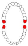
Maxillary (Upper) Molars
Maxillary molars are the large teeth located at the back of the upper jaw. There are six of them in total, with three on each side: the first, second, and third molars, named from the front of the mouth to the back. These teeth are crucial for chewing food and maintaining the vertical dimension of the face.
General Features of Maxillary Molars
- Not Succedaneous: Unlike most other teeth, molars do not replace any primary (baby) teeth.
- Wider Buccolingually: They are broader from side to side (buccolingually) than from front to back (mesiodistally).
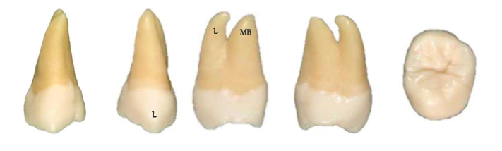
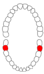
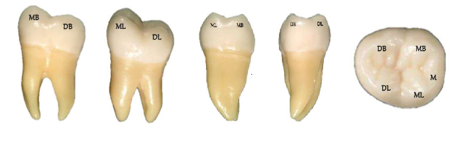
Maxillary First Molar
Overview
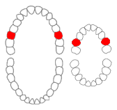
The maxillary first molar is the largest tooth in the upper jaw and the first of the maxillary molars. They typically have four main cusps and one additional minor cusp.
All teeth are given teeth numbers for identification purposes. For the purposes of this article the universal teeth numbering system, the American teeth numbering system will be used. There is also the World Dental Federation numbering system. In adults, the maxillary first molars are teeth number 3 (right tooth) and 14 (left tooth). In children, these are teeth B (right tooth) and I (left tooth).
Adult upper first molars typically erupt in the mouth (grow into your mouth) at the age of 6-7, while baby upper first molars erupt between 13-19 months.
Defining Features
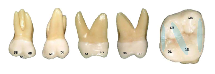
- Buccal View (Cheek Side): Shows two roots, with a third root visible from other angles.
- Lingual View (Tongue Side): Displays the larger cusp (humps on the teeth) and could have a unique cusp of Carabelli visible here.
- Occlusal View (Top View): Features a rhomboidal shape with an oblique ridge running diagonally across the tooth. The grooves of this tooth can form an H or Y pattern.
Maxillary Second Molar
Overview
The second molar is slightly smaller than the first molar and typically has four cusps arranged in a less complex pattern.
In adults, the maxillary second molars are teeth number 2 (right tooth) and 15 (left tooth). In children, these are teeth A (right tooth) and J (left tooth).
Adult upper second molars typically erupt in the mouth at the age of 12-13, while baby upper second molars erupt between 25-33 months.
Defining Features
Differences from the first molar include:
- Shorter Crown (Whiter Part): The crown is shorter vertically (occlusocervically).
- Narrower Roots: The roots are narrower and less divergent.
- Occlusal View (Top View): Shows a more rectangular or rhomboidal shape with a simpler groove pattern, often resembling an H.
Maxillary Third Molar
Overview
The third molar, or wisdom tooth, varies greatly in shape and size but generally is smaller than the first and second molars. Notable characteristics include:
In adults, the maxillary third molars are teeth number 1 (right tooth) and 16 (left tooth). They typically erupt between the ages of 17-21. Children do not have wisdom teeth.
Defining Features
- Highly Variable Tooth: Often resembles the first or second molar but is generally smaller and more irregular.
- Roots: Shorter, usually fused together, and significantly curved.
- Occlusal View (Top View): Highly irregular patterns.
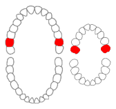
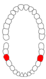
Mandibular Molars
Mandibular molars are the large teeth located at the back of the lower jaw. There are six of them in total, with three on each side: the first, second, and third molars, named from the front of the mouth to the back. These teeth play a crucial role in chewing food and maintaining the vertical dimension of the face.
General Features of Mandibular Molars
- Not Succedaneous: Unlike most other teeth, molars do not replace any primary (baby) teeth.
- Wider Mesiodistally: They are broader from front to back (mesiodistally) than from side to side (buccolingually).
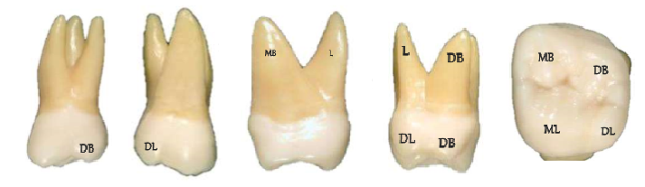
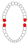
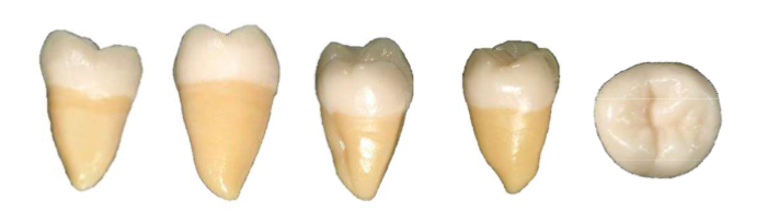
Mandibular First Molar
The mandibular first molar is the largest tooth in the lower jaw. It typically has five cusps: three on the buccal (cheek) side and two on the lingual (tongue) side. Key features include:
In adults, the mandibular first molars are teeth number 30 (right tooth) and 19 (left tooth). In children, these are teeth S (right tooth) and L (left tooth).
Adult lower first molars typically erupt in the mouth at the age of 6-7, while baby lower first molars erupt between 14-18 months.
Defining Features
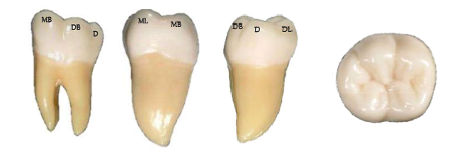
- Buccal View (Cheek Side): Shows the mesial and distal roots, with the mesial root being straighter initially but curving at the end.
- Occlusal View (Top View): Features a Y-shaped groove pattern and three primary fossae: central, mesial triangular, and distal triangular.
Mandibular Second Molar
Overview
The second molar, slightly smaller than the first, typically has four cusps arranged in a “+” pattern.
In adults, the mandibular second molars are teeth number 31 (right tooth) and 18 (left tooth). In children, these are teeth S (right tooth) and L (left tooth).
Adult lower second molars typically erupt in the mouth at the age of 11-13, while baby lower second molars erupt between 23-31 months.
Defining Features
- Shorter Crown (Whiter Part): The crown is shorter vertically (occlusocervically).
- Narrower Roots: The roots are narrower and more inclined.
- Occlusal View (Top View): Shows a “+” groove pattern formed by the central, lingual, and buccal grooves.
Mandibular Third Molar
Overview
The third molar, or wisdom tooth, varies greatly in shape and size but generally is smaller than the first and second molars.
In adults, the mandibular third molars are teeth number 32 (right tooth) and 17 (left tooth). They typically erupt between the ages of 17-21. Children do not have wisdom teeth.
Defining Features
- Highly Variable Crown: Often resembles the first or second molar but is generally smaller and more irregular.
- Roots: Shorter, usually fused together, and significantly curved.
- Occlusal View: A highly irregular pattern.
Anatomy of Molar Teeth
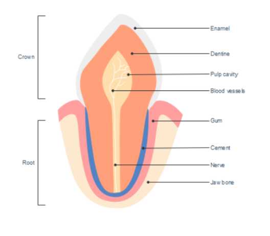
Like all teeth, molars have two main parts: the crown and the root.
- Crown: The crown is the part of the tooth that is visible in the mouth. As discussed previously, molars have a flatter chewing surface on their crowns, known as an occlusal surface
- Root: The root is the part of the tooth that is embedded in the jawbone. molars typically have 2-3 roots, while teeth such as incisors can have only one.
Each tooth is composed of three main parts: enamel, dentin, and pulp.
- Enamel: The enamel is the hardest part of the tooth and is what gives the tooth its white color. It is also the hardest substance in the human body. Its function is to protect the tooth from chewing and biting forces.
- Dentin: Dentin is a hard, yellowish material that makes up the majority of the tooth. Its function is to support the enamel and protect the pulp from bacteria.
- Pulp: The pulp is the innermost part of the tooth that contains blood vessels and nerves. Its function is to provide nutrients and sensation to the tooth.
How to Keep Your Teeth Clean
Your molars , just like every other tooth, are vital to proper oral health, and it is important to take care of them! Not cleaning them properly can lead to cavities, gum disease, and eventually tooth loss.
- Brush your teeth twice a day with fluoride toothpaste: Flouride is a mineral that helps to remineralize your teeth and prevent cavities. Be sure to brush all teeth surfaces, including near the gum line, and pay attention to your back teeth. The most frequently missed locations.
- Floss ATLEAST every day (But recommended twice a day): Flossing removes plaque and germs from between your teeth, helping to prevent cavities between teeth. Be sure to hug the curve of each tooth as you floss and go slightly under the gumline.
- Visit the dentist every six months: Regular dental visits are important in order to catch any problems early and to keep your teeth healthy! They can also provide cleanings that are more thorough than what you can do at home.
Disclaimer
The contents of this website, such as text, graphics, images, and other material are for informational purposes only and are not intended to be substituted for professional medical advice, diagnosis, or treatment. Nothing on this website constitutes the practice of medicine, law or any other regulated profession.
No two mouths are the same, and each oral situation is unique. As such, it isn’t possible to give comprehensive advice or diagnose oral conditions based on articles alone. The best way to ensure you’re getting the best dental care possible is to visit a dentist in person for an examination and consultation.
SAVE TIME AND MONEY AT ANY DENTIST
Less dental work is healthier for you. Learn what you can do to minimize the cost of dental procedures and avoid the dentist altogether!
About the Reviewer
Dr. Adam Szymczak is a general dentist and owner of Smile Care Dental with over 20 years of dental experience, focusing on implant, reconstructive, asleep and surgical dentistry. Upon graduation, he was hired by Mount Sinai Hospital in Toronto, where he completed the highly sought-after postgraduate general practice residency program receiving training in multi-disciplinary treatment, sedation, surgery, and implant dentistry.