

Wisdom teeth, also known as third molars, are the last set of teeth to emerge in the mouth. These teeth typically appear during the late teens or early twenties, long after the other permanent teeth have erupted. While some individuals have healthy wisdom teeth, others experience complications that necessitate removal. To provide you with a clearer understanding of wisdom teeth and their locations in the mouth, we have compiled a collection of real pictures outside of the mouth, pictures inside the mouth, and X-Rays taken at dental offices nationwide. By studying these images, you can gain a better understanding of how wisdom teeth appear, as well as the common difficulties associated with their removal.
What are Your Wisdom Teeth?
The wisdom teeth, or third molars, are the last set of permanent teeth to emerge in the mouth. In dental terminology, the process of these teeth coming into the mouth is called “erupting.” Typically, these wisdom teeth begin to erupt around 17-21 years old. Like other adult molars, they are used for chewing and grinding food. This means their shape and structure are often bulkier looking than the teeth closer to the front of the mouth, namely the incisors and canines.
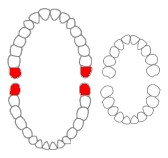
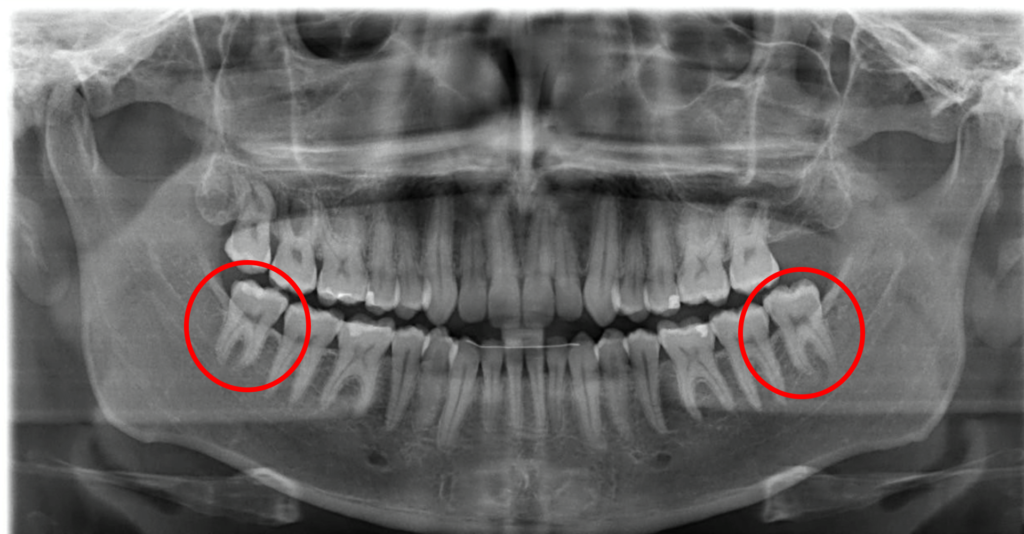
There are usually four wisdom teeth in each corner of your mouth – two in the upper jaw (maxillary wisdom teeth) and two in the lower jaw (mandibular wisdom teeth). However, fewer (or no wisdom teeth) or more than four wisdom teeth may sometimes be present. In the picture above, the wisdom teeth are highlighted in red.
All teeth are given teeth numbers for identification purposes. For the purposes of this article, the universal teeth numbering system, the American teeth numbering system, will be used. There is also the World Dental Federation numbering system. The maxillary wisdom teeth are numbered 1 and 16. The mandibular wisdom teeth are numbered 17 and 32.
Wisdom teeth can sometimes cause dental problems due to their size and location. If the teeth do not have room to erupt fully, they may become impacted or “stuck” beneath the gums. This can lead to pain, swelling, infection, and even damage to the surrounding teeth and gums. Sometimes, wisdom teeth may require surgical removal to prevent further complications.
Pictures of Wisdom Teeth: What do they Look Like?
Truthfully, wisdom teeth are the teeth that often appear the most different from person to person. Sometimes they might not even look like teeth at all. This is because a lot of their growth in the mouth will be dictated by the teeth around it and the space available to grow in their jaw. Here is what some typical wisdom teeth look like outside of the mouth.
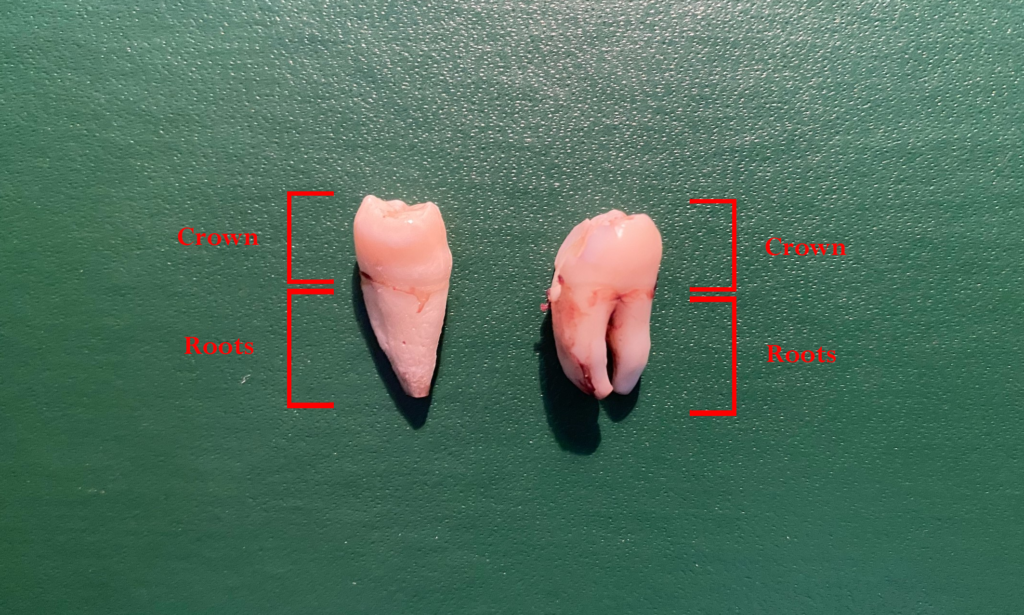
Wisdom teeth are composed of two main parts: the crown and the root.
Crown
The crown is the part of the tooth visible in the mouth. Surprisingly, wisdom teeth typically have the smallest crown of all your back teeth (the molars). They are usually rectangular and bulky and shape, perfect grinding. On the occlusal surface of these teeth (the tooth surface you bite on), these teeth typically have three to five cusps (bumps) that help grind food.
Root
The root is the part of the tooth embedded in the jawbone. Wisdom teeth typically have 2-3 roots, making them more difficult to remove than other teeth. These roots are usually shorter than the other molars, especially if they are removed from the mouth before completely growing. Because of the lack of space in the jaw, it is completely normal to have roots that are fused (stuck) together.
Each tooth is further composed of three main parts: enamel, dentin, and pulp.
- Enamel: The enamel is the hardest part of the tooth and is what gives the tooth its white color. It is also the hardest substance in the human body. Its function is to protect the tooth from chewing and biting forces.
- Dentin: Dentin is a hard, yellowish material that makes up the majority of the tooth. Its function is to support the enamel and protect the pulp from bacteria.
- Pulp: The pulp is the innermost part of the tooth that contains blood vessels and nerves. Its function is to provide nutrients and sensation to the tooth.
Real X-Ray Pictures of Wisdom Teeth
When trying to understand wisdom teeth, it is important to not only see pictures of them but also look at X-ray pictures. Inside the mouth, it is normal to see three types of wisdom teeth: fully-impacted wisdom teeth, partially-impacted wisdom teeth, and non-impacted wisdom teeth.
Fully Impacted Wisdom Teeth
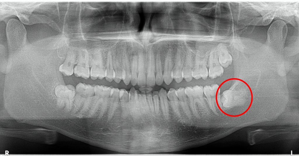
Fully-impacted wisdom teeth are those that are unable to erupt through the gums in your mouth (and may remain entirely inside the jawbone). They can be completely vertical in the mouth and are not visible to the naked eye. Most people do not have wisdom teeth that are fully impacted, but for those who do, it is possible to avoid wisdom teeth removal altogether. Keeping these wisdom teeth can actually have a few benefits. This is because no bacteria can enter the tooth and cause decay, making it very unlikely for a cavity to form.
Partially Impacted Wisdom Teeth
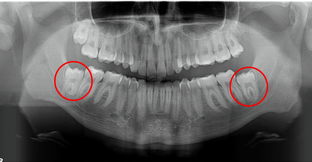
Partially-impacted wisdom teeth are teeth that have partially grown into the mouth. This means they have broken partially through the jawbone and/or partially through the gum tissue. Most people will have partially impacted wisdom teeth. They may be angled either horizontally or vertically, but they cannot erupt fully due to a lack of space in the jaw. These teeth can be more difficult to keep clean and often require surgical removal for proper dental hygiene.
Non-Impacted Wisdom Teeth (Erupted Wisdom Teeth)
Non-impacted wisdom teeth are another uncommon type of wisdom teeth. These are the teeth that can fully erupt on their own without any intervention from a dentist or surgeon. They look just like any other molar and have similar features to regular molars. These teeth are likely not to cause any problems if they are properly cared for and cleaned. However, many people are unable to brush far enough back in their mouth to clean these teeth properly, leading to an increased risk of gum disease and cavities.
Conclusion
These real pictures of wisdom teeth provide valuable insights into their appearance, conditions, and complications. While not all wisdom teeth are problematic, the majority often require extraction due to tooth decay. If you are uncertain about the state of your wisdom teeth, it is best to consult your dentist for a thorough evaluation.
Disclaimer
The contents of this website, such as text, graphics, images, and other material are for informational purposes only and are not intended to be substituted for professional medical advice, diagnosis, or treatment. Nothing on this website constitutes the practice of medicine, law or any other regulated profession.
No two mouths are the same, and each oral situation is unique. As such, it isn’t possible to give comprehensive advice or diagnose oral conditions based on articles alone. The best way to ensure you’re getting the best dental care possible is to visit a dentist in person for an examination and consultation.
SAVE TIME AND MONEY AT ANY DENTIST
Less dental work is healthier for you. Learn what you can do to minimize the cost of dental procedures and avoid the dentist altogether!