

Tooth abscesses can be a serious dental condition that develops from untreated tooth decay. Understanding the stages of a tooth abscess is crucial for timely treatment. In this article, we will discuss the stages of tooth decay leading up to a tooth abscess, along with corresponding pictures, symptoms, and treatment options. It is important to note that no two teeth are unique. Therefore, one picture may not accurately depict the stage of abscess on your own tooth. For example, these stages may be visible on an X-Ray but not visually apparent in a picture or the other way around.
Stage Zero – Tooth Decay: Cavities
Tooth decay, or cavities, is the initial stage preceding tooth abscess. Cavities start as small areas of decay on the enamel, the outermost layer of the tooth. Over time, decay can progress through the dentin and reach the innermost layer, the pulp. As decay progresses, it can cause the tooth to become painful and sensitive to hot or cold temperatures. However, if this decay is allowed to progress to an abscess, the severity of the discomfort may become unbearable. This is why prevention, through brushing, flossing, and eating less sugary foods, is key when it comes to good oral hygiene. The stages of tooth decay are:
Enamel Decay
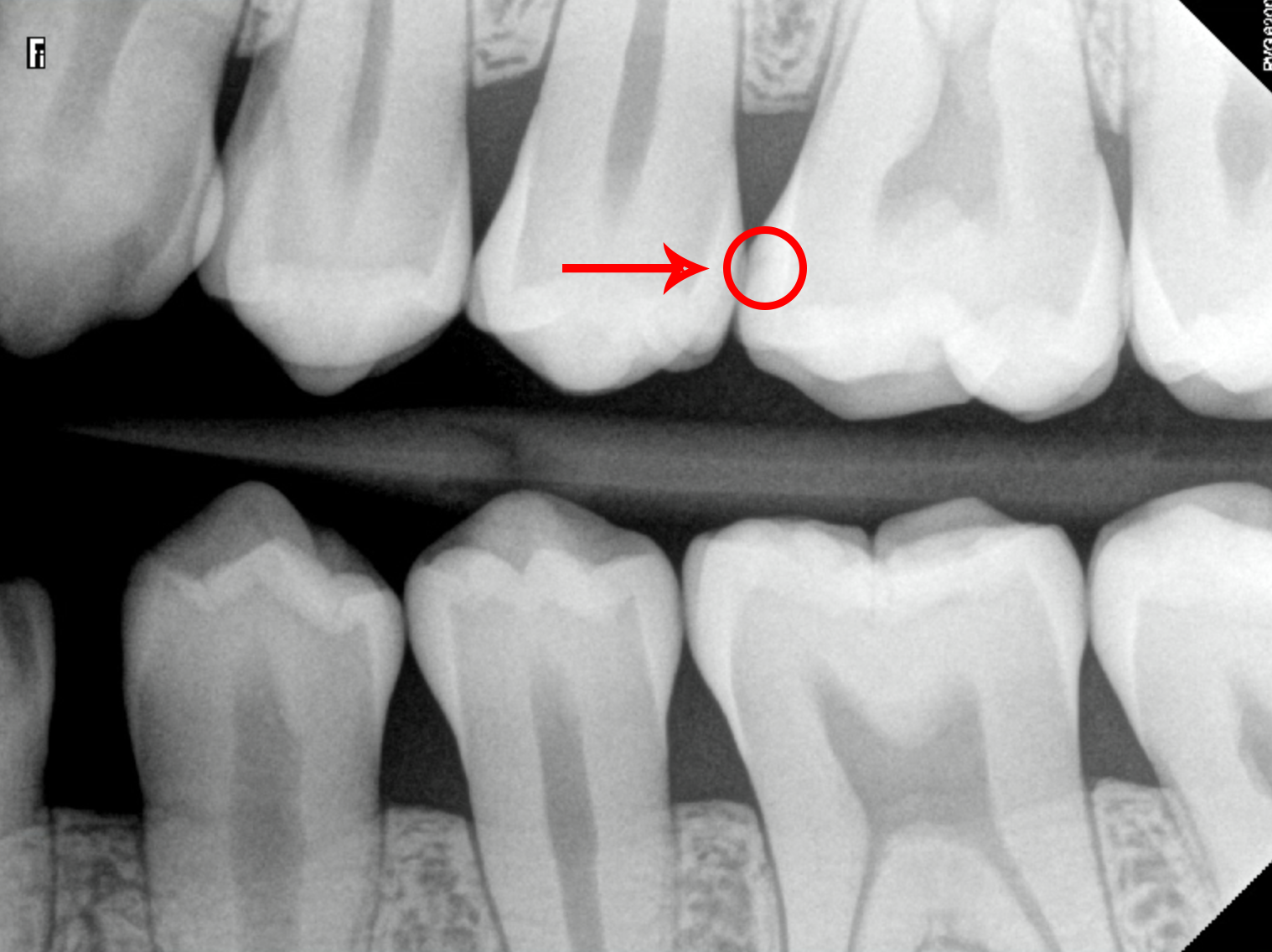
Enamel decay occurs when the cavity has only broken down your tooth’s strong outer layer (enamel).
- Appearance: To the eye, it typically looks like small brown or black spots on the tooth and may feel soft and sticky. X-Rays of cavities typically show small black spots, known as radiolucencies, which may be visible.
- Symptoms: These cavities are usually painless and not sensitive, making them difficult to detect without a dental check-up.
- Treatment: A small tooth filling will typically be performed, sometimes without local anesthesia. In cases where the cavity is caught extremely early, the cavity is also reversible via fluoride treatment.
Dentin Decay
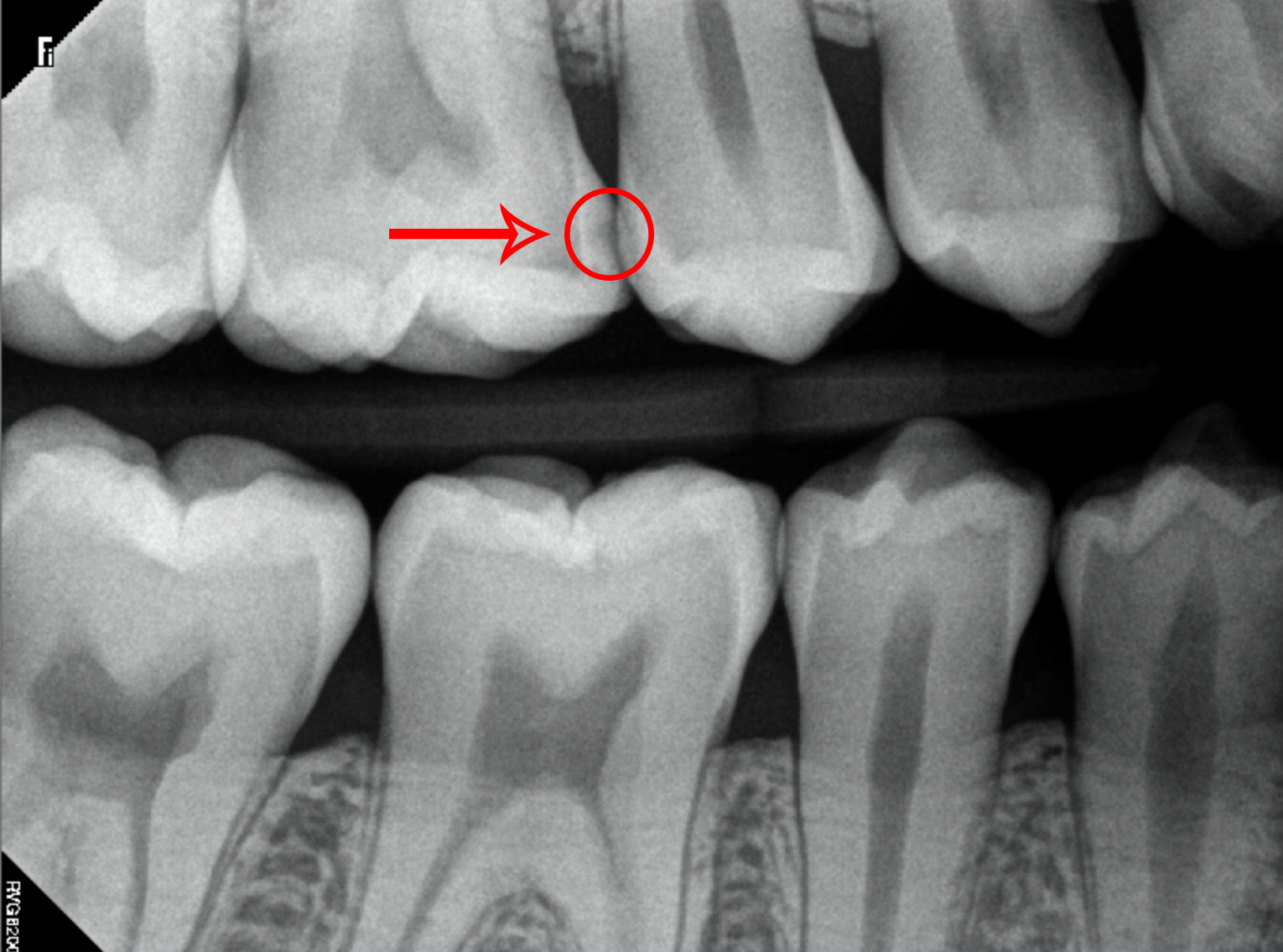
Dentin decay occurs when the cavity has broken through the enamel and entered the second softer layer of your tooth (dentin).
- Appearance: Larger brown or black spots may be visible to the eye, possibly with a hole forming. On an X-Ray, radiolucencies become bigger and wider.
- Symptoms: Patients will often complain of sensitivity to sweet foods and hot or cold temperatures, but not always and not persistently. The cavity might also feel soft and mushy and will progress quickly due to its location in the second softer layer of the tooth.
- Treatment: A small to medium-sized filling is typically performed with local anesthesia. Extra precautions may be taken to ensure proper restoration if these cavities are close to the nerve or the gum line.
Pulp Decay
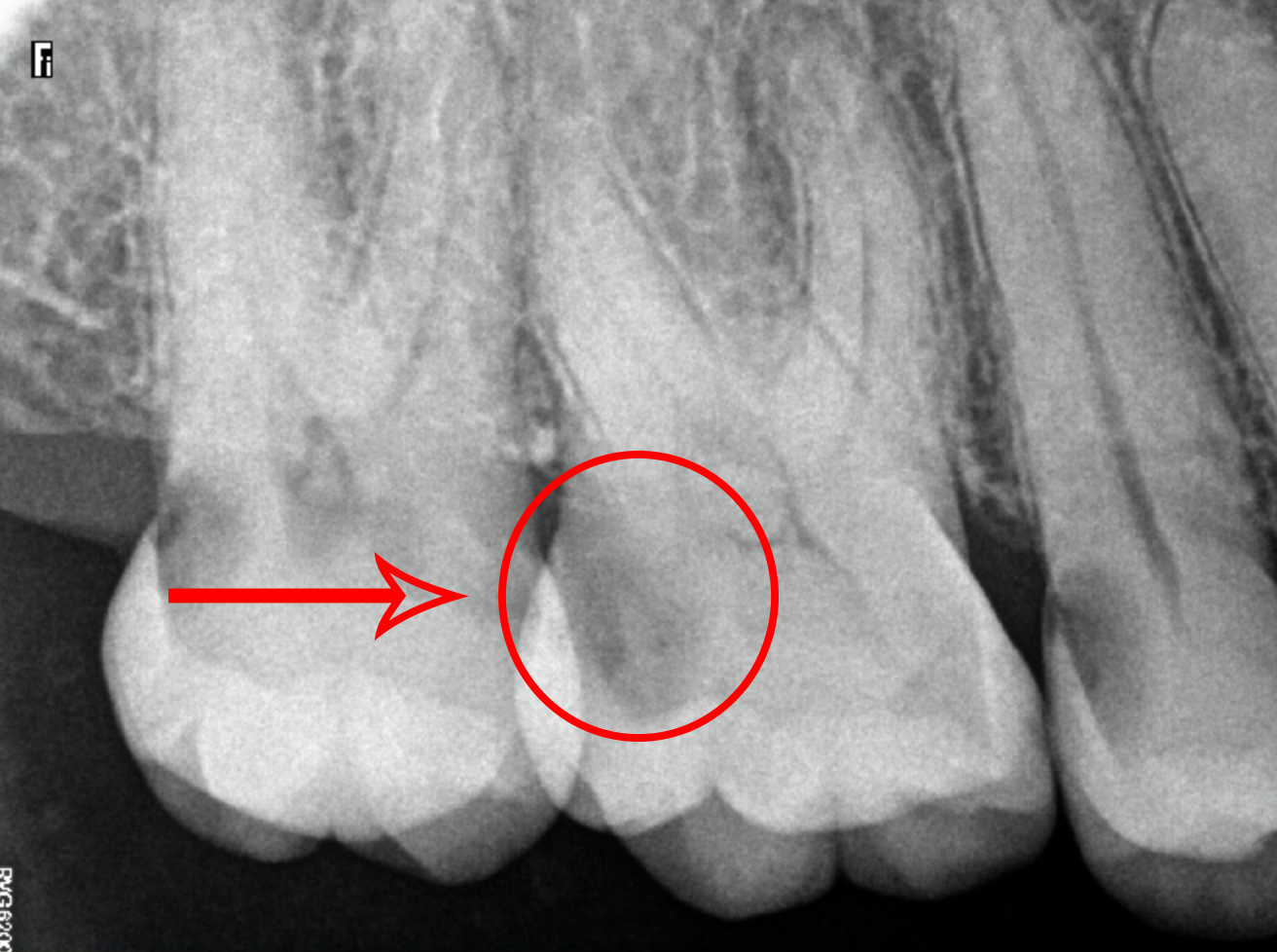
Pulp decay occurs when the cavity has broken through the dentin and entered the center of your tooth (pulp).
- Appearance: Pulp decay looks like a large brown or black hole in the tooth. On an X-Ray, radiolucencies become bigger and wider and can spread from one side of the tooth to the other.
- Symptoms: Unbearable toothache or throbbing pain due to the involvement of the nerve located in the inner portion of your tooth.
- Treatment: A root canal is typically required to treat the infected nerve while preserving the tooth. In extreme cases, the tooth may need to be extracted and later replaced with an implant, crown, or bridge.
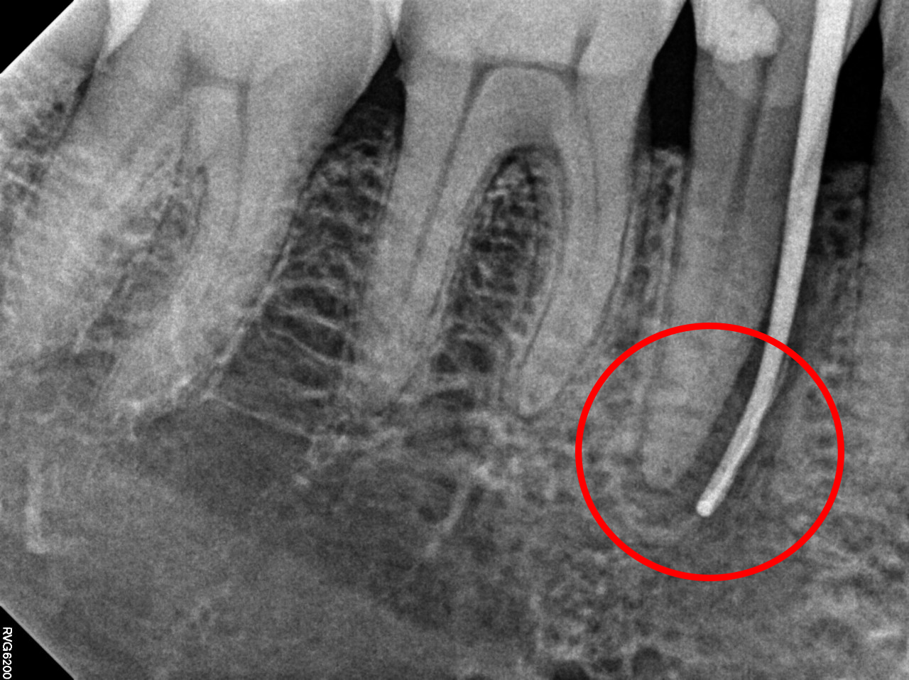
Stage One – Periapical Pathology (PAP): Initial Abscess Formation
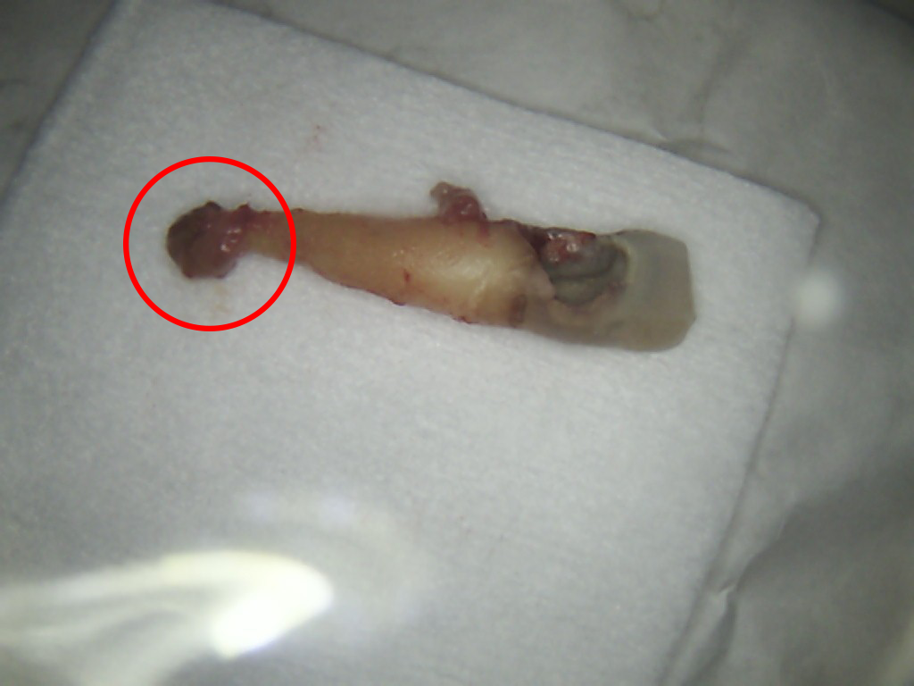

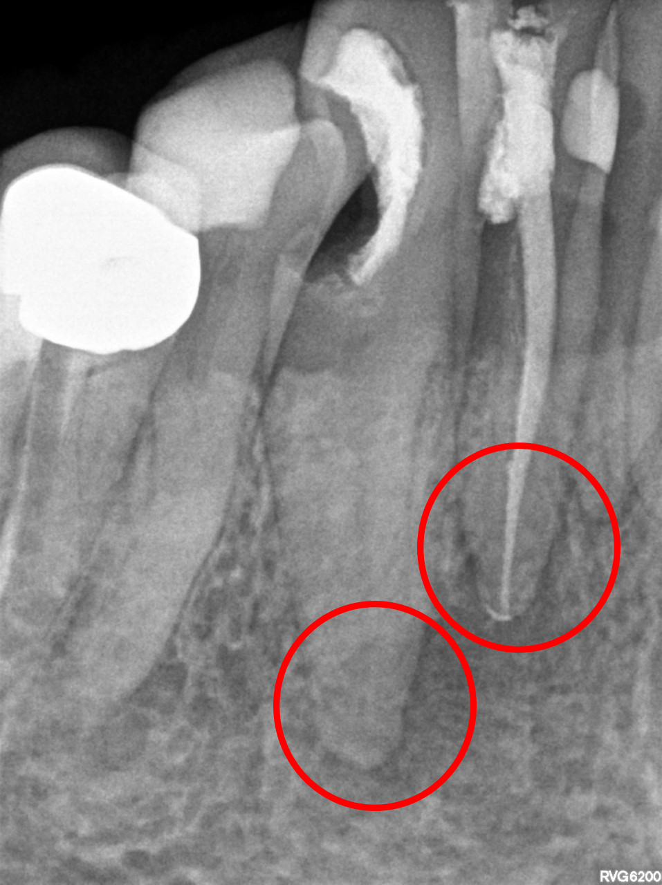
In this stage, the infection spreads through the entirety of the pulp to the tips of the root of the teeth, resulting in what is known as a periapical pathology. This is when a tooth infection has begun to spread out of the tip of your tooth’s roots and into the surrounding bone.
- Appearance: Periapical Pathology is usually hidden within the bone and only visible after removing it from the tooth. A dental X-ray typically reveals a darker circle at the tips of your teeth.
- Symptoms: A toothache and throbbing pain can be felt in the area surrounding the abscessed tooth.
- Treatment: Early stage may be treated with a root canal with antibacterial medication being taken before completing the root canal. In extreme cases, the tooth may need to be extracted and later replaced with an implant, crown, or bridge.
Stage Two – Parulis (Abscess on the Gum)
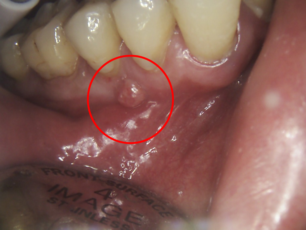
At this stage, the infection penetrates the bone and forms a pimple on the gums, known as a parulis or gum boil.
- Appearance: Pimple-like swelling on the gums, usually pink or red in color, containing pus.
- Symptoms: Surprisingly, there is typically less pain in this stage than in the initial abscess formation. This is because the puss that has built up in the jaw and caused pain has found a way to escape through the creation of the abscess.
- Treatment: Typically, a root canal, antibiotics, and drainage of the gum boil are necessary. In extreme cases, the tooth may need to be extracted and later replaced with an implant, crown, or bridge.
Stage Three – Facial Swelling

Facial swelling occurs as the untreated infection progresses. This is due to the continuous production of pus and bacteria accumulating locally, often leading to a fever.
- Appearance: Swelling on one side of the face, increasing in size each day.
- Symptoms: Tender, warm, and disfiguring swelling, often resembling a golf ball in the cheeks.
- Treatment: Drainage of the swelling, a full course of antibiotics, and subsequent treatment of the infected tooth with a root canal or extraction will be required.
Stage Four – Airway Compromise
This critical stage occurs when swelling extends to the throat, where the trachea is located. This is where oxygen is brought into the lungs, and carbon dioxide is brought out of . If this stage is not treated properly and in a timely manner, airway obstruction can occur. This can be life-threatening, resulting in death. DO NOT DELAY TREATMENT.
- Appearance: Massive swelling of the throat and lower jaw, similar to having a baseball stuck in the area.
- Symptoms: Excruciating pain, difficulty breathing, talking, and eating.
- Treatment: Seek immediate medical help at a hospital, as it requires specialized care, drainage, and antibiotics.
Conclusion
Prevention is the best strategy for avoiding a tooth abscess. Regular brushing, flossing, and regular dental check-ups can help keep your teeth healthy and strong. If you are experiencing any symptoms of a tooth abscess, it’s important to seek treatment as soon as possible. If left untreated, the infection can spread and even become life-threatening. Seeing pictures of the different tooth abscess stages can help you identify if you are suffering from a tooth abscess and get prompt treatment. Furthermore, it is important to speak with a qualified dental professional if you suspect that you have a tooth abscess to ensure that you are getting the best possible care.
Disclaimer
The contents of this website, such as text, graphics, images, and other material are for informational purposes only and are not intended to be substituted for professional medical advice, diagnosis, or treatment. Nothing on this website constitutes the practice of medicine, law or any other regulated profession.
No two mouths are the same, and each oral situation is unique. As such, it isn’t possible to give comprehensive advice or diagnose oral conditions based on articles alone. The best way to ensure you’re getting the best dental care possible is to visit a dentist in person for an examination and consultation.
SAVE TIME AND MONEY AT ANY DENTIST

Less dental work is healthier for you. Learn what you can do to minimize the cost of dental procedures and avoid the dentist altogether!