

The temporalis muscle is one of the four muscles of mastication, which are primarily responsible for the movements of the jaw during chewing and speaking. The temporalis muscle is notable for its fan-shaped appearance and is a crucial component in the elevation and retraction of the mandible. Understanding the anatomy, function, and clinical significance of the temporalis muscle can provide valuable insights into its role in both normal mastication and potential disorders of the temporomandibular joint (TMJ).
| Key Facts | Details |
|---|---|
| Origin | Bony surfaces of the temporal fossa, inferior temporal line, and overlying temporal fascia. |
| Insertion | Medial aspect of the coronoid process of the mandible and anterior margin of the mandibular ramus, extending almost to the last molar tooth. |
| Action | Elevation of the mandible (closing the mouth), retraction of the mandible (pulling the jaw backward), and maintaining the resting position of the mandible. |
| Innervation | Deep temporal nerves, branches of the mandibular nerve (cranial nerve V3). |
| Blood Supply | Deep temporal arteries (branches of the maxillary artery) and anastomoses with branches of the middle temporal artery. |
Anatomy of the Temporalis Muscle
Location and Structure
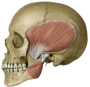
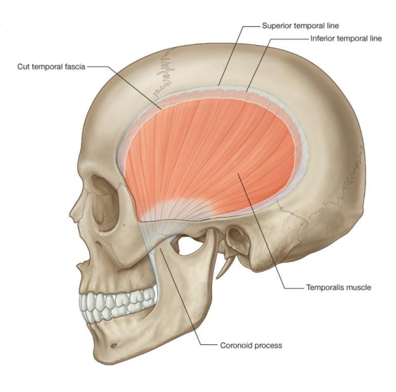
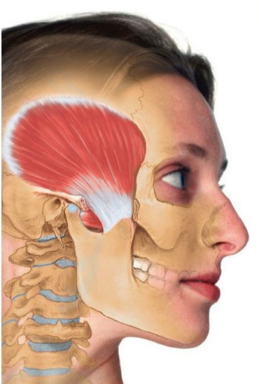
The temporalis muscle is a broad, fan-shaped muscle that occupies the temporal fossa, a shallow depression on the side of the skull. This muscle is covered by a strong layer of connective tissue known as the temporal fascia, which helps anchor the muscle to the skull.
Origin and Insertion
- Origin: The temporalis muscle originates from the bony surfaces of the temporal fossa and the inferior temporal line of the skull. It also attaches to the overlying temporal fascia, which provides additional structural support.
- Insertion: The muscle fibers converge to form a strong tendon that passes deep to the zygomatic arch (the bony prominence of the cheek). This tendon inserts into the medial aspect of the coronoid process of the mandible and the anterior margin of the mandibular ramus, extending almost to the last molar tooth.
Orientation of Muscle Fibers
The muscle fibers of the temporalis are arranged in different directions, allowing for a range of movements:
- Anterior Fibers: These fibers are oriented vertically and are primarily responsible for elevating the mandible (closing the mouth).
- Posterior Fibers: These fibers are more horizontal and play a significant role in retracting the mandible (pulling the jaw backward).
Function of the Temporalis Muscle
The temporalis muscle is involved in several key movements of the mandible:
- Elevation of the Mandible: The primary function of the temporalis muscle is to elevate the mandible, which is essential for closing the mouth. This action is primarily performed by the vertical (anterior) fibers of the muscle.
- Retraction of the Mandible: The posterior fibers of the temporalis muscle are responsible for retracting the mandible, pulling it backward. This movement is important for adjusting the position of the jaw during chewing and speaking.
- Resting Tonus: The temporalis muscle maintains a resting tonus, which helps keep the mandible in its normal resting position when the mouth is closed but not actively clenching.
- Lateral Movements: The temporalis muscle also contributes to the lateral (side-to-side) movements of the mandible, which are necessary for the grinding motion during chewing.
Innervation and Blood Supply
Innervation
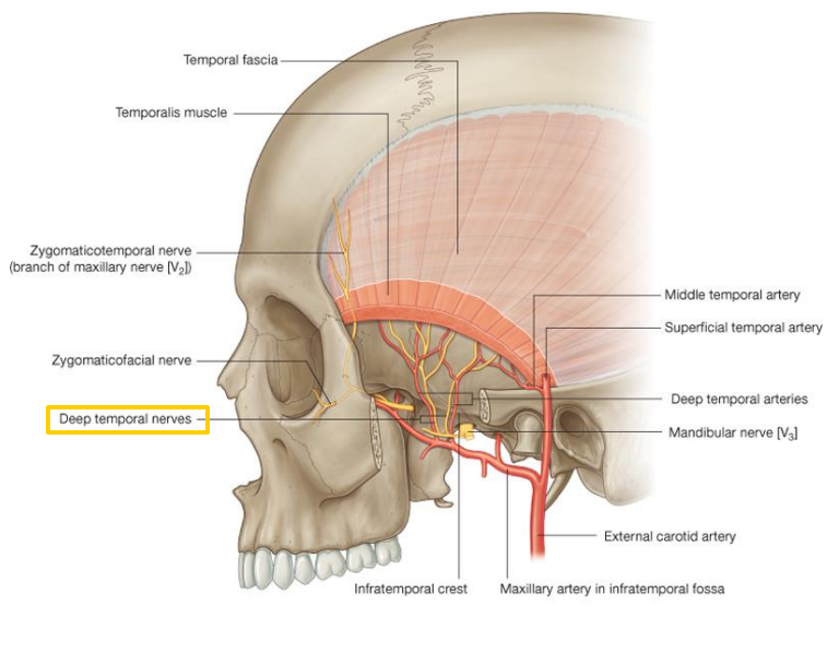
The temporalis muscle is innervated by the deep temporal nerves, which are branches of the mandibular nerve (cranial nerve V3). These nerves provide the motor signals necessary for muscle contraction.
Blood Supply
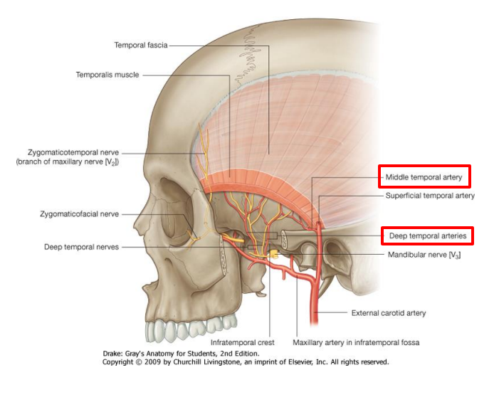
The blood supply to the temporalis muscle comes from the deep temporal arteries, which are branches of the maxillary artery. These arteries travel alongside the deep temporal nerves and provide the necessary oxygen and nutrients to the muscle. The deep temporal arteries also anastomose (connect) with branches of the middle temporal artery, which further supports the vascularization of the muscle.
Clinical Significance
Temporomandibular Joint Disorders
The temporalis muscle plays a significant role in the function of the temporomandibular joint (TMJ). Dysfunction or pain in the temporalis muscle can contribute to TMJ disorders, which are characterized by pain and compromised movement of the jaw. Common symptoms of TMJ disorders include:
- Jaw Pain: Pain in the jaw, which can radiate to the ear, temple, or neck.
- Difficulty in Chewing: Pain or discomfort during chewing or other jaw movements.
- Jaw Clicking or Popping: Audible clicking or popping sounds when opening or closing the mouth.
- Limited Jaw Movement: Difficulty in fully opening or closing the mouth.
Myofascial Pain Syndrome
Myofascial pain syndrome (MPS) is a chronic pain condition that can affect the temporalis muscle. It is characterized by the presence of trigger points, which are sensitive areas within the muscle that can cause referred pain. Treatment for MPS may include physical therapy, massage, and medications to alleviate pain and reduce muscle tension.
Conclusion
The temporalis muscle is a critical component of the masticatory system, contributing to the elevation, retraction, and lateral movements of the mandible. Its complex structure and orientation allow for a wide range of jaw movements necessary for chewing and speaking. Understanding the anatomy and function of the temporalis muscle is essential for diagnosing and treating disorders related to the temporomandibular joint and masticatory system. If you experience any symptoms related to jaw pain or dysfunction, it is important to consult with a healthcare professional for proper evaluation and management.
Disclaimer
The contents of this website, such as text, graphics, images, and other material are for informational purposes only and are not intended to be substituted for professional medical advice, diagnosis, or treatment. Nothing on this website constitutes the practice of medicine, law or any other regulated profession.
No two mouths are the same, and each oral situation is unique. As such, it isn’t possible to give comprehensive advice or diagnose oral conditions based on articles alone. The best way to ensure you’re getting the best dental care possible is to visit a dentist in person for an examination and consultation.
SAVE TIME AND MONEY AT ANY DENTIST

Less dental work is healthier for you. Learn what you can do to minimize the cost of dental procedures and avoid the dentist altogether!