

The muscles of mastication are a group of four muscles that are primarily responsible for the movement of the jaw. These muscles are essential for chewing (mastication), speaking, and any other activity that involves the opening and closing of the mouth. The muscles of mastication are located in the parotid and infratemporal regions of the head, and they work in harmony to allow for complex movements of the jaw. This article gives an overview of each muscle of mastication, their function, insertion and origin, and blood and nerve supplies.
Anatomy and Function of the Muscles of Mastication
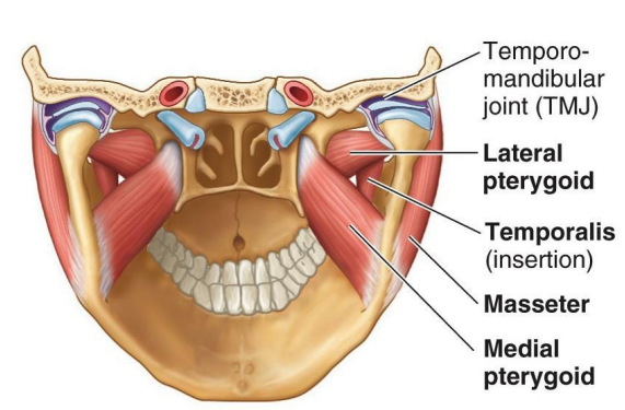
The four main muscles of mastication are the temporalis, masseter, medial pterygoid, and lateral pterygoid muscles. Each muscle has distinct anatomical features and functions, contributing to various movements of the mandible (lower jaw).
Temporalis Muscle
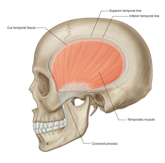
The temporalis muscle is a large, fan-shaped muscle that fills the temporal fossa on the side of the skull. It is covered by a strong temporal fascia, which helps anchor the muscle to the skull.
- Origin: The temporalis muscle originates from the bony surfaces of the temporal fossa and the inferior temporal line.
- Insertion: It inserts into the medial aspect of the coronoid process of the mandible and the anterior margin of the mandibular ramus.
Function: The temporalis muscle is responsible for elevating the mandible (closing the mouth) and retracting the mandible (pulling the jaw backward). Its anterior fibers are oriented vertically, making them ideal for elevation, while the posterior fibers are more horizontal, aiding in retraction.
Innervation and Blood Supply: The muscle is innervated by the deep temporal nerves, which are branches of the mandibular nerve (CN V3). The deep temporal arteries, branches of the maxillary artery, supply blood to the muscle.
Masseter Muscle
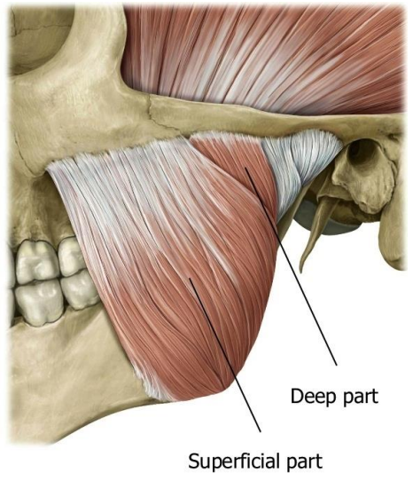
The masseter muscle is one of the most powerful muscles involved in chewing. It is quadrangular in shape and covers the lateral aspect of the mandibular ramus.
- Superficial Head:
- Origin: Maxillary process of the zygomatic bone and the anterior two-thirds of the zygomatic arch.
- Insertion: Angle of the mandible and the lateral surface of the mandibular ramus.
- Deep Head:
- Origin: Medial aspect of the zygomatic arch and the posterior third of the zygomatic arch.
- Insertion: Central and upper parts of the mandibular ramus, as high as the coronoid process.
Function: The masseter muscle elevates the mandible (closing the mouth) and assists in protrusion (pushing the jaw forward) and retrusion (pulling the jaw backward). Its deep fibers are involved in retrusion, while the superficial fibers assist in protrusion.
Innervation and Blood Supply: The masseteric nerve, a branch of the mandibular nerve (CN V3), innervates the muscle. Blood supply comes from the masseteric artery, a branch of the maxillary artery, and contributions from the transverse facial artery.
Medial Pterygoid Muscle
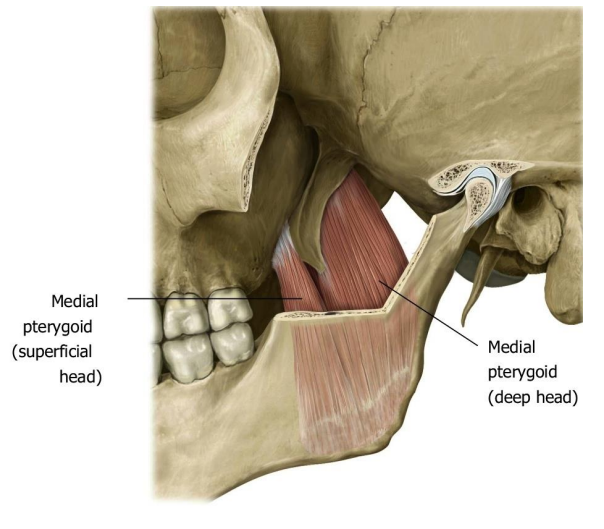
The medial pterygoid muscle is quadrangular and has two heads: a deep head and a superficial head. It lies on the medial aspect of the mandibular ramus, almost mirroring the masseter muscle.
- Superficial Head:
- Origin: Tuberosity of the maxilla and the pyramidal process of the palatine bone.
- Insertion: Pterygoid rugosity on the medial aspect of the mandibular ramus near the angle.
- Deep Head:
- Origin: Medial surface of the lateral plate of the pterygoid process.
- Insertion: Joins the superficial head to insert on the pterygoid rugosity on the medial aspect of the mandibular ramus.
Function: The medial pterygoid muscle elevates the mandible (closing the mouth) and assists in protrusion. It also contributes to lateral (side-to-side) movements of the mandible, helping in the grinding motion during chewing.
Innervation and Blood Supply: The nerve to the medial pterygoid, a branch of the mandibular nerve (CN V3), innervates the muscle. Blood supply comes from the pterygoid branches of the maxillary artery.
Lateral Pterygoid Muscle
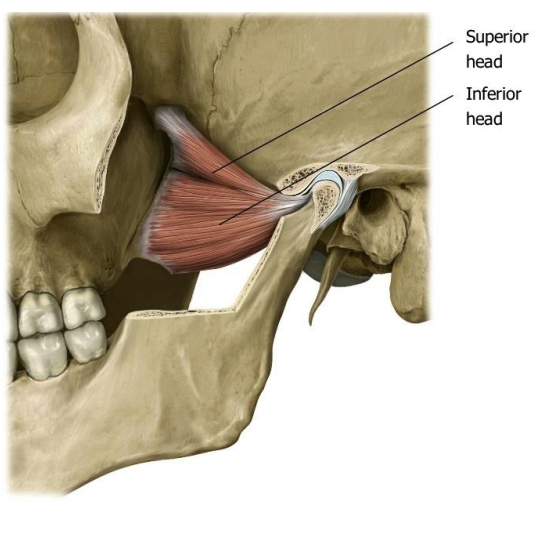
The lateral pterygoid muscle is a triangular muscle located within the infratemporal fossa. It is the only muscle of mastication that is oriented horizontally and has two heads: a superior (upper) head and an inferior (lower) head.
- Superior Head:
- Origin: Roof of the infratemporal fossa (greater wing of the sphenoid bone).
- Insertion: Capsule of the temporomandibular joint (TMJ) and the articular disc.
- Inferior Head:
- Origin: Lateral aspect of the lateral pterygoid plate.
- Insertion: Pterygoid fovea on the neck of the mandibular condyle.
Function: The lateral pterygoid muscle is primarily responsible for protracting the mandible (pushing the jaw forward). It also assists in mandibular depression (opening the mouth) and plays a role in lateral (side-to-side) movements. The superior head is particularly active during the power stroke of chewing (closure on resistance).
Innervation and Blood Supply: The nerve to the lateral pterygoid, a branch of the mandibular nerve (CN V3), innervates the muscle. Blood supply is provided by the pterygoid branches of the maxillary artery.
Coordinated Movements of the Jaw
The muscles of mastication work together to perform the complex movements required for chewing and other jaw functions. Here is how they coordinate:
- Elevation (Closing the Mouth): The temporalis, masseter, and medial pterygoid muscles contract to elevate the mandible.
- Depression (Opening the Mouth): The lateral pterygoid muscles and the suprahyoid muscles (located in the neck) assist in opening the mouth.
- Protrusion (Pushing the Jaw Forward): The lateral pterygoid, medial pterygoid, and superficial head of the masseter work together to push the mandible forward.
- Retrusion (Pulling the Jaw Backward): The posterior fibers of the temporalis and the deep head of the masseter pull the mandible backward.
- Lateral Movements (Chewing): The masseter and temporalis muscles on one side, along with the medial and lateral pterygoid muscles on the opposite side, allow for side-to-side grinding movements during chewing.
Importance in Dental Health
The proper function of the muscles of mastication is crucial for effective chewing and overall oral health. Dysfunction or pain in these muscles can lead to temporomandibular joint (TMJ) disorders, which can cause significant discomfort and difficulty in jaw movement. If you experience any pain or difficulty while chewing or moving your jaw, it is important to consult with a dental professional who can evaluate the health and function of your muscles of mastication.
Disclaimer
The contents of this website, such as text, graphics, images, and other material are for informational purposes only and are not intended to be substituted for professional medical advice, diagnosis, or treatment. Nothing on this website constitutes the practice of medicine, law or any other regulated profession.
No two mouths are the same, and each oral situation is unique. As such, it isn’t possible to give comprehensive advice or diagnose oral conditions based on articles alone. The best way to ensure you’re getting the best dental care possible is to visit a dentist in person for an examination and consultation.
SAVE TIME AND MONEY AT ANY DENTIST
Less dental work is healthier for you. Learn what you can do to minimize the cost of dental procedures and avoid the dentist altogether!