
Lateral Pterygoid Muscle
The lateral pterygoid muscle is a key player in the movements of the mandible, specifically in opening the mouth and moving the jaw side-to-side. This article provides a comprehensive overview of the lateral pterygoid muscle, detailing its anatomy, function, and clinical significance.
| Aspect | Details |
|---|---|
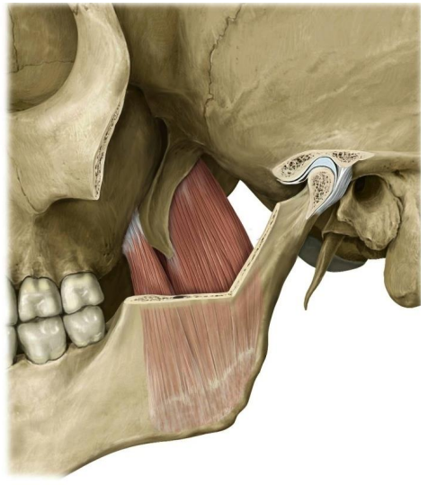
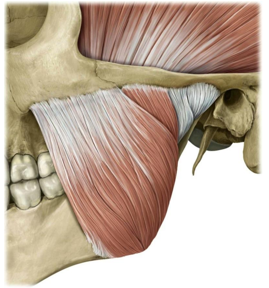
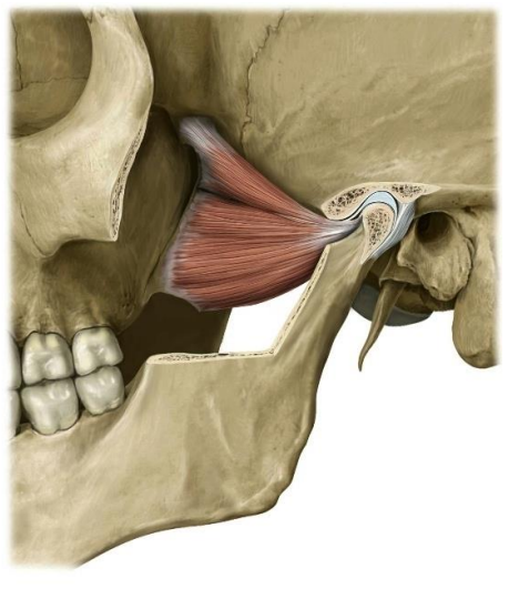
| Origin | Superior head: Roof of the infratemporal fossa (greater wing of the sphenoid and infratemporal crest). Inferior head: Lateral surface of the lateral pterygoid plate. |
| Insertion | Superior head: Capsule of the TMJ and articular disc. Inferior head: Pterygoid fovea on the neck of the mandible. |
| Action | Bilateral: Protraction of the mandible, assists in depression (opening the mouth). Unilateral: Contralateral excursion (lateral movement). |
| Innervation | Nerve to the lateral pterygoid, a branch of the mandibular nerve (cranial nerve V3). |
| Blood Supply | Pterygoid branches of the maxillary artery. |
0 Comments
July 13, 2024