

When people hear the word “cyst”, they often imagine something painful or dangerous. But in dentistry, odontogenic cysts—cysts that arise from tissues involved in tooth development—are more common and usually less scary than they sound.
This blog will help you understand what these cysts are, how dentists identify them, and what treatments look like.
What Is an Odontogenic Cyst?

An odontogenic cyst is essentially a small, fluid-filled sac that forms in your jaw or gums. These cysts develop from the same cells that help form your teeth.
They are:
- Pathologic cavities (meaning they shouldn’t be there)
- Filled with fluid
- Lined with epithelium (a thin tissue layer)
Most odontogenic cysts are benign—non-cancerous—but they still require attention to prevent complications.
What Do These Cysts Feel Like?
Surprisingly, most cysts are painless unless they become infected or very large. That’s why many cysts are discovered by accident during routine dental X-rays.
Some may feel like:
- A firm, painless swelling
- A slight bump inside the mouth
- A bluish soft swelling (especially eruption cysts in children)
How Dentists Detect Cysts
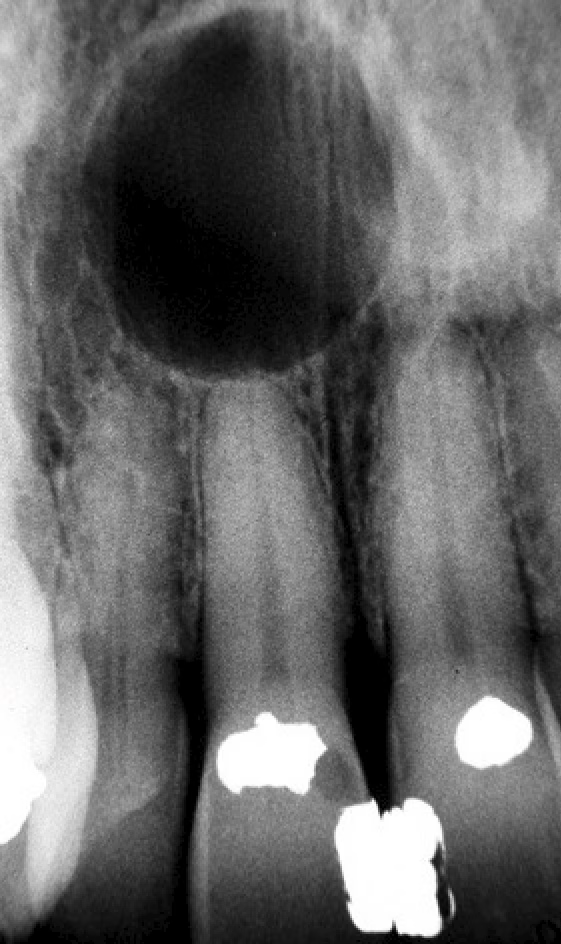
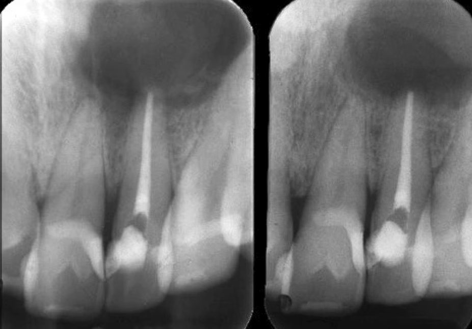
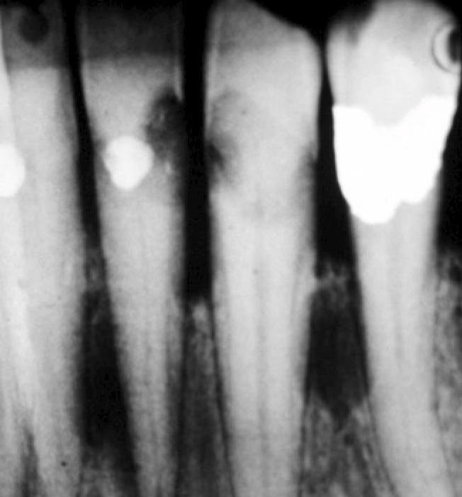
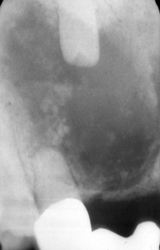
Dentists rely heavily on radiographs (X-rays) and 3D scans to diagnose cysts. Cysts often look like:
- A dark circular area on an X-ray (called a radiolucency)
- Well-defined edges, sometimes with a thin white border
- Changes in the surrounding structures, such as:
- Teeth shifting out of place
- Bone expanding
- Thinning of the jawbone walls
- Displacement of important anatomy (like the mandibular nerve canal)
Because many cysts grow quietly, regular dental X-rays help catch them early.
Types of Odontogenic Cysts
Dentists classify these cysts into two major groups: inflammatory and developmental.
1. Inflammatory Odontogenic Cysts
Radicular Cyst (Most Common)
This is the most common cyst dentists see.
It forms when the pulp (nerve) of a tooth dies and inflammation spreads to the tip of the root, triggering cyst formation.
Key facts:
- Always associated with a non-vital (dead) tooth
- Usually not painful
- Treated by addressing both the tooth and the cyst (root canal or extraction + cyst removal)
You may also hear the term apical granuloma—a related condition that looks similar on X-rays.
Residual Cyst
This cyst is simply a radicular cyst left behind after a tooth was removed.
It has the same features but appears in an area where a tooth used to be.
2. Developmental Odontogenic Cysts
These cysts form because of how teeth develop—not because of infection.
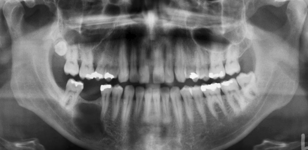
Dentigerous Cyst
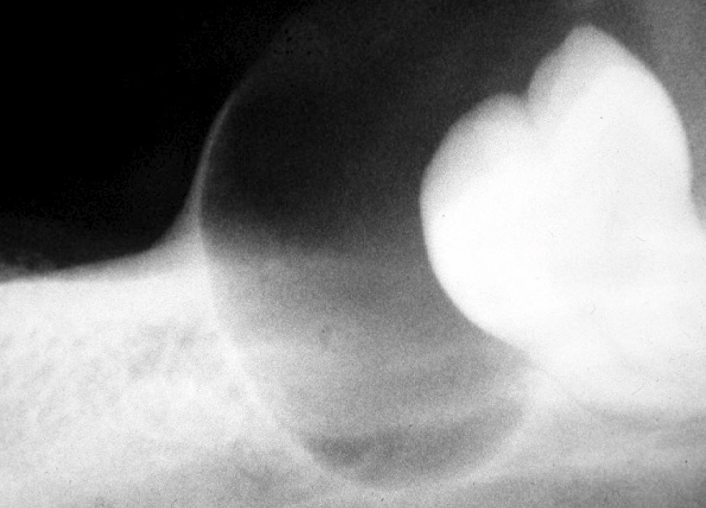
The second most common odontogenic cyst.
It forms around the crown of an unerupted tooth, usually wisdom teeth or canines.
Identifying features:
- Surrounds an unerupted tooth’s crown
- Attached at the tooth’s cemento-enamel junction (CEJ)
- May cause bone expansion or tooth displacement
Eruption Cyst

Common in young children.
This is the soft-tissue version of a dentigerous cyst.
It appears as a bluish, soft swelling over an erupting tooth.
Most of the time, it resolves on its own as the tooth breaks through the gums.
Odontogenic Keratocyst (OKC)
Previously called a keratocystic odontogenic tumour (KOT), an OKC is unique because of its:
- High recurrence rate
- Tendency to grow extensively before symptoms appear
- Location (often in the back of the lower jaw)
It has a very characteristic microscopic appearance, including a thin, keratin-producing lining.
Lateral Periodontal Cyst
This cyst forms along the side of a vital tooth root, often in the lower premolar area.
A variant called a botryoid odontogenic cyst can have multiple compartments.
Calcifying Odontogenic Cyst (Gorlin Cyst)
A rare cyst that may contain calcifications (small “hard spots”) and special cells called ghost cells.
Found mostly in the front teeth area, and sometimes associated with other dental tumors.
Buccal Bifurcation Cyst
A cyst occurring on the cheek side (buccal aspect) of the first or second lower molar in children.
Key features:
- Affects vital teeth
- Causes buccal swelling
- Usually treated without removing the tooth
How Are These Cysts Treated?
Treatment varies depending on the type, size, and location of the cyst. Common methods include:
✔️ Enucleation
Surgical removal of the entire cyst.
✔️ Curettage
Scraping the area to remove residual cyst cells.
✔️ Marsupialization
Creating an opening to allow the cyst to shrink before full removal—often used for large cysts.
✔️ Follow-Up
Essential because some cysts, like OKCs, can come back.
Why Should You Care About Odontogenic Cysts?
Even though many cysts are painless, untreated cysts can:
- Weaken the jawbone
- Shift or loosen teeth
- Delay tooth eruption in children
- Damage nerves
- Get infected
- Become very large before detection
Routine dental checkups and X-rays make early diagnosis and treatment easy and predictable.
Final Thoughts
Odontogenic cysts are common, and most are harmless when found and treated early. If your dentist notices a cyst on an X-ray, don’t panic—these conditions are well-understood, manageable, and often treated with simple outpatient procedures.
If you have concerns about a cyst or dental swelling, talk to your dentist or an oral surgeon. Early assessment is the best step toward protecting your oral health.
Disclaimer
The contents of this website, such as text, graphics, images, and other material are for informational purposes only and are not intended to be substituted for professional medical advice, diagnosis, or treatment. Nothing on this website constitutes the practice of medicine, law or any other regulated profession.
No two mouths are the same, and each oral situation is unique. As such, it isn’t possible to give comprehensive advice or diagnose oral conditions based on articles alone. The best way to ensure you’re getting the best dental care possible is to visit a dentist in person for an examination and consultation.
SAVE TIME AND MONEY AT ANY DENTIST
Less dental work is healthier for you. Learn what you can do to minimize the cost of dental procedures and avoid the dentist altogether!